The most common and best documented stricture is PRAA. However, there are a few others that have appeared as well in our ME dogs. They are listed below:

Unusual vascular ring anomaly associated with a persistent right aortic arch in two dogs:
House AK1, Summerfield NJ, German AJ, Noble PJ, Ibarrola P, Brockman DJ.
Author information
https://www.ncbi.nlm.nih.gov/pubmed/1635573
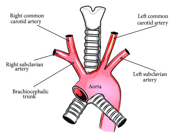
Abstract: An unusual vascular ring anomaly consisting of a persistent right aortic arch and a left ligamentum arteriosum extending from the main pulmonary artery to an aberrant left subclavian artery and left aortic arch remnant complex was identified in a German shepherd dog and a great Dane. The left subclavian artery and left aortic arch remnant complex originated at the junction between the right distal aortic arch and the descending aorta and coursed dorsal to the oesophagus in a cranial direction. The attachment of the ligamentum arteriosum to the aberrant left subclavian artery was approximately 5 cm cranial to the point of origin of the aberrant left subclavian artery and left aortic arch remnant complex from the descending aorta in both dogs. This anomaly observed in both dogs is similar to an anomaly reported in humans, in which a persistent right aortic arch is found in conjunction with an aberrant left subclavian artery and a left aortic arch remnant (Kommerell’s diverticulum). Surgical ligation and division of the left ligamentum arteriosum in both dogs, along with division of the left subclavian artery in the great Dane, resulted in resolution of clinical signs in both of the dogs in this report.
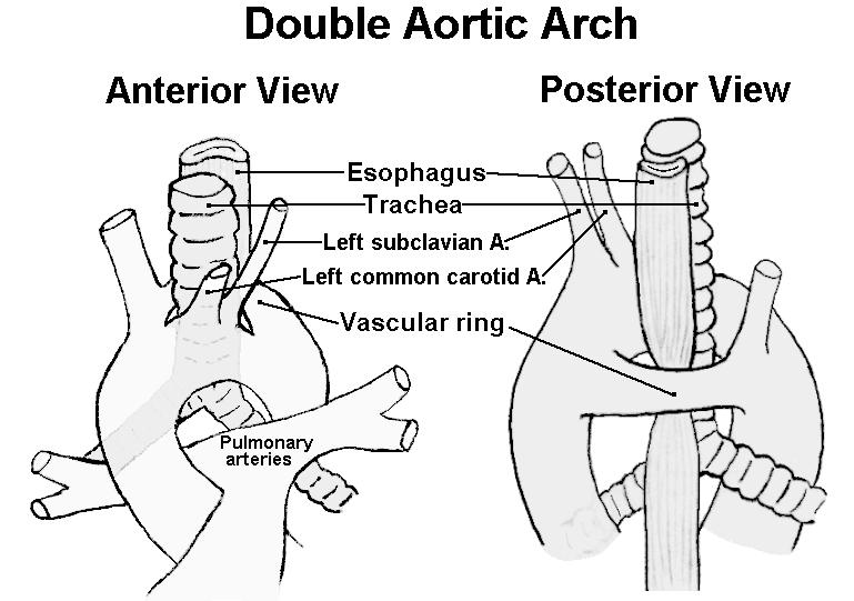
Double aortic arch in a dog:
Article in Journal of the American Veterinary Medical Association 225(8):1222-4, 1196-7 · November 2004 with 20 Reads
DOI: 10.2460/javma.2004.225.1222 · Source: PubMed
https://www.researchgate.net/publication/8199948_Double_aortic_arch_in_a_dog
Vascular ring anomalies are developmental anomalies of the thoracic great vessels resulting in complete or partial encircling of the esophagus and the trachea by a vascular ring formation. Persistent right aortic arch with left ligamentum arteriosum accounts for 95% of vascular ring anomalies in dogs. The dog in this report had a double aortic arch, which is a type 4 vascular ring anomaly. Double aortic arch is a rare congenital heart defect resulting from the improper development of the embryonic arches. The prognosis for dogs that have undergone surgery for correction of double aortic arches is generally regarded as poor. The dog in this report underwent surgery for correction of double aortic arches and recovered without dilation or motility disorders of the esophagus. Results indicate that small animals that undergo early surgical correction of double aortic arches with relief of esophageal constriction can have a good prognosis. To the authors’ knowledge, there have been no previous reports of dogs that have survived long enough to be discharged from the hospital after surgical correction of double aortic arches.